Bird Vision – A World to Discover
News
Why focus on bird vision ?
The visual acuity of the eagle or of the swallow diving on its prey and catching it in flight is amazing. Most of the so-called modern birds, or Neoaves, have a similar or, for some species, even a finer vision than ours.
The bird eye may help us to better understand how our one is built and how it works. It also offers an opportunity to ask ourselves about the causes of different eye diseases, such as glaucoma or age-related macular degeneration (AMD).
Learning more about bird vision
Bird vision is outstanding
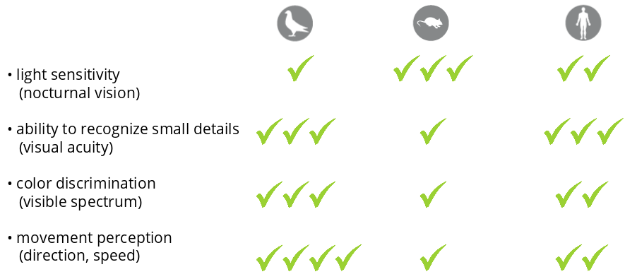
Visual performance depends on four distinct properties : 1) sharp perception (or visual acuity), 2) color discrimination, 3) movement perception (speed, orientation, direction), 4) light sensitivity. The first two properties are in primates much superior to those of other mammals but similar to those of modern birds. On the other hand, birds such as passerines, raptors or columbines are more skillful than primates in detecting rapid movements.
Why the diurnal vision of birds and primates has no equivalent in the animal world ?
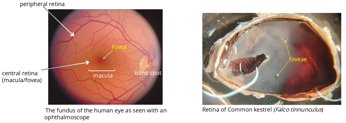
Based on our current knowledge, it is thought to be related to the presence of a specialized region of the retina that is called the fovea. Among mammals, only primates have a fovea. Although the retinas of fewer than a hundred bird species (1% of the total number of species) have been studied to date, the vast majority of diurnal modern birds (Neoaves) are thought to have one or, for some of them, two foveae. On the other hand, fowls (hens, ducks, geese) have no fovea at all. The emergence of the fovea in birds during evolution has contributed to improve the flight.
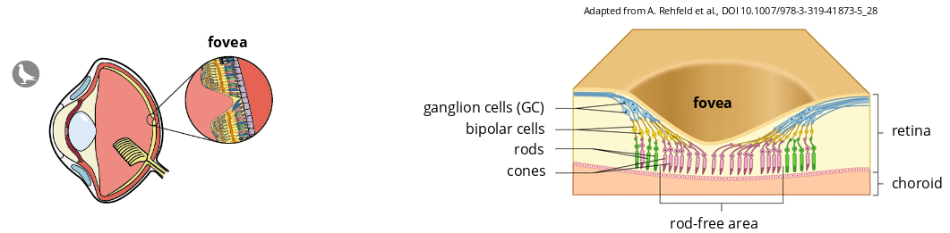
What is the fovea ?
The fovea is a specialized region of the retina. It displays specific properties that greatly influence our perception of colors and image sharpness.
Transduction of visual input at the level of the fovea
In the retina, photoreceptors (cone and rods) transform light signals into electrical impulses which are then transmitted to Ganglion Cells (GCs). These retinal neurons transmit visual input via the optic nerve to different regions of the brain.
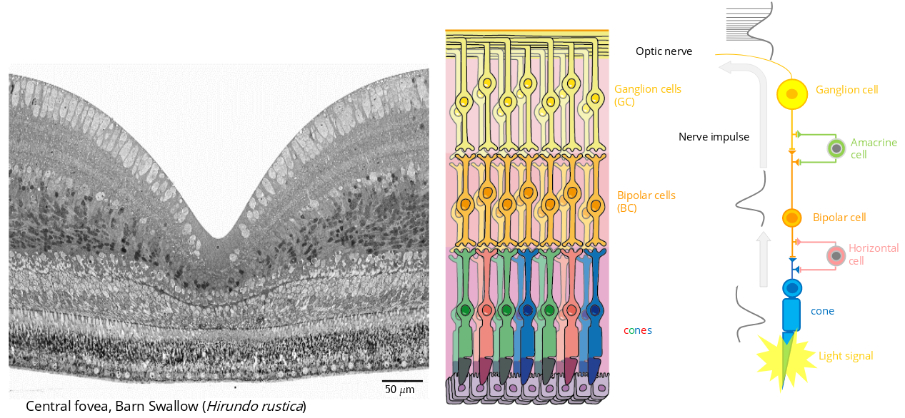
Why does the fovea have exceptional properties ?
In the fovea, a single Ganglion Cell (GC) receives signals produced by a single cone. Likewise, the receptive field of one GC corresponds to the width of one cone. In contrast, outside the fovea, a single GC makes connection with several rods, and the area occupied by these rods define the receptive field of the GC. Therefore, the receptive fields of GC are, in general, smaller inside than outside the fovea. There is an analogy between the size of a receptive field and that of a pixel. Smaller pixels often means better image quality. Likewise, images transmitted to the brain by the fovea will be perceived by the brain as sharper than those from retinal neurons located outside the fovea.

Foveal vision and evolution
Recent genomic analyzes suggest that modern birds, or Neoaves, split from their ancestors, the Galloanserae, at the time of the Cretaceous-Paleogene extinction event 66 million years ago. This evolutionary branch of birds was so successful that several thousand new species have appeared within a relatively short period of time. These modern birds have colonized lands and seas all over the world. We do not know the origin of this exceptional success story. One hypothesis that we put forward would be a better spatio-temporal visual acuity resulting from the appearance of foveal vision. A marked increase in visual acuity undoubtedly improved the quality of flight, which became faster, more precise and more sustained. The conquest of the sky by Neoaves had major consequences on their metabolism and body shape, making it stronger and fitter. Improved flight and increased visual acuity also favored dietary changes. While Galloanserae are predominantly vegetarian and ground-feeding, Neoaves feed primarily on live prey including insects, crustaceans, worms and small vertebrates.
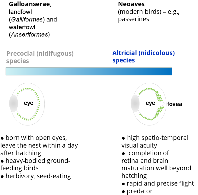
We found that the presence of a fovea in the retina is accompanied by significant changes in the organization of the region of the brain named the optic tectum. Increased complexity of retinal and brain tissues requires the lengthening of development well beyond hatching. Unlike precocial chicks which are self-sufficient immediately after hatching and quickly leave the nest, the large majority of Neoaves chicks are born with closed eyes and cared for by their parents for several weeks. This shift from an early mode of development to a development that extends beyond hatching has required dramatic changes in behavior. The extended stay of the chicks in the nest gave the brain time to develop new functions. In a nutshell, the Neoaves have become smarter than their ancestors.
Why focus on the fovea ?
With very high neuron densities and intense activity, the fovea is a region of the nervous system of highest energy demands. In humans, the fovea and the surrounding retinal area, named the macula, can be the target of retinal diseases such as the infamous age-related macular degeneration (AMD). This neurodegenerative disease affects a growing number of our fellow citizens. The importance of the fovea for vision and its susceptibility to eye diseases should encourage us to intensify research in this area.
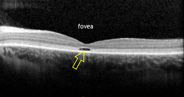
Another example of human eye disease : macular dystrophy
A macular dystrophy highlights the singular character of the fovea. The authors in Vaclavik et al., 2013 reported two cases of patients with a neurodegenerative disease called spinocerebellar ataxias (SCA) and associated with a loss of vision (acuity and colors). This disease is caused by a mutated gene (sca1) that encodes an abnormal protein called ATXN1. These patients display a foveal cavity corresponding to the specific loss of cones of the fovea.
Research projects
The fovea in humans and birds was discovered at the end of the 19th century, but the scattered studies that followed did not allow significant advances in our knowledge of this retinal specialty. The fovea is a complex neural tissue and the tools needed to study its structure and function were not available until recently. This lack of knowledge is also linked to the fact that most of the research in visual science and ophthalmology are carried out in mice and zebrafish, two species that have no macula and no fovea. Non-human primates would be the best models for studying the fovea, but their access to experimentation is limited for obvious ethical reasons. Recently, the development of organoids which reproduce in vitro the micro-anatomy of an organ like the eye has aroused much interest. Unfortunately, research conducted to date has failed to observe the development of a fovea in retina-like organoids derived from human pluripotent stem cells.
Currently, three ongoing research projects are supported by the Foundation Gene&Vision.ch :